Data Utilization
This is a model that estimates the age from fundus images, created using data from health checkup facilities collected by the Japan Ocular Imaging Registry. While its performance has been validated in internal verification, it does not guarantee its performance with external data. The model is intended to be used in ophthalmology research that utilizes fundus images.
The model is available for free use, and the Japan Ophthalmological Society, Japanese Society of Artificial Intelligence in Ophthalmology, Japan Ocular Imaging Registry, and National Institute of Informatics do not claim ownership of any outcomes resulting from the use of this model. The Japan Ophthalmological Society, Japanese Society of Artificial Intelligence in Ophthalmology, Japan Ocular Imaging Registry, and National Institute of Informatics are not responsible for any output or other results generated by the model.
If you use this model, please cite the following paper and acknowledge the Japan Ophthalmological Society, Japan Ocular Imaging Registry, and National Institute of Informatics in your acknowledgments.
[Citation]
Miyake M, Akiyama M, Kashiwagi K, Sakamoto T, Oshika T. Japan Ocular Imaging Registry: a national ophthalmology real-world database. Jpn J Ophthalmol. 2022 Nov;66(6):499-503. doi: 10.1007/s10384-022-00941-0. Epub 2022 Sep 23. PMID: 36138192.
[Acknowledgment Example]
"The pre-training model used in this study was developed by the Japanese Ophthalmological Society and the National Institute of Informatics, and was made available through the Japan Ocular Imaging Registry website(http://www.joir.jp/)."
Retinal images and health examination data were collected from a single health examination facility. The dataset consisted of 12,734 images from 12,734 individuals (7,375 right eyes and 5,359 left eyes) who had no abnormalities in their retinas and were “healthy”*. The left eye images were flipped horizontally and used for training as if they were all right eye images.
* "healthy" was defined as meeting the following conditions:
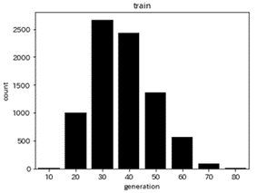
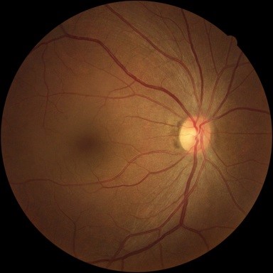
Out of 12,734 retinal images, we used 8,149 images for training, 2,038 for validation, and 2,547 for testing. The actual age of the training data was 42.2 ± 11.3 years (minimum 18 years, maximum 84 years) and the distribution is shown in Figure 1. We created an AI model by transfer learning using the Swin Transformer model1 trained on Imagenet2, with the PyTorch library3 in Python34. The input image size was set to 384×384, and an example of an input image is shown in Figure 2.
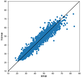
The actual age of the test data was 42.3 ± 11.0 years. The scatter plot of actual and predicted ages is shown in Figure 3. The mean absolute error (MAE) between actual age and age predicted by the AI model was 2.39 years.
| gender | male | female |
|---|---|---|
| people | 3474 | 4675 |
| age | 10s | 20s | 30s | 40s | 50s | 60s | 70s | 80s |
|---|---|---|---|---|---|---|---|---|
| people | 11 | 1004 | 2665 | 2432 | 1366 | 569 | 91 | 11 |
The AI model has been made available in the pth format, and a sample of its operation has been described separately in the source code. Python3 and Pytorch environments are required for operation.
This model predicts gender from fundus images using data collected from health check-up facilities as part of the Japan Ocular Imaging Registry. While the model has demonstrated the performance described below during internal validation, its performance with external data is not guaranteed. It is intended for use in research on gender-related diseases.
This model is freely available for use. Any outcomes derived from the use of this model do not belong to the Japanese Ophthalmological Society, the Japan Society for Ophthalmic AI, the Japan Ocular Imaging Registry (JOIR), nor the National Institute of Informatics. These organizations do not assume any responsibility for the outputs or other results generated by this model.
When utilizing this model, please cite the following paper and acknowledge the Japanese Ophthalmological Society, the Japan Ocular Imaging Registry, and the National Institute of Informatics in the Acknowledgments section.
[Citation]
Miyake M, Akiyama M, Kashiwagi K, Sakamoto T, Oshika T. Japan Ocular Imaging Registry: a national ophthalmology real-world database. Jpn J Ophthalmol. 2022 Nov;66(6):499-503. doi: 10.1007/s10384-022-00941-0. Epub 2022 Sep 23. PMID: 36138192.
[Example Acknowledgment]
"The pre-training model used in this study was developed by the Japanese Ophthalmological Society and the National Institute of Informatics, and was made available through the Japan Ocular Imaging Registry website(http://www.joir.jp/)."
This model was developed using 163,789 fundus images and accompanying health check-up data collected from a single health check-up facility.
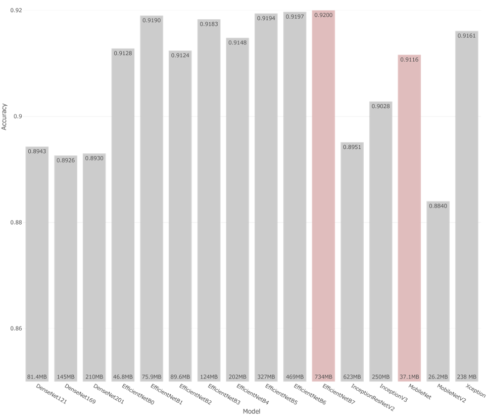
Of the 163,789 fundus images, 80% were used for training and 20% for testing. The training dataset consisted of 56.1% male and 43.9% female images, while the test dataset contained 56.4% male and 43.6% female images. AI models were developed using 16 architectures (DenseNet-121/169/201, Inception-v3, Inception-ResNet-v2, MobileNet, MobileNetV2, Xception, EfficientNet-B0/B1/B2/B3/B4/B5/B6/B7) trained with the TensorFlow library in Python 3. The input image size was standardized to 384×384 pixels. Examples of input images are shown in Figure 1.
When using the AI models to predict gender from test dataset fundus images, the highest accuracy achieved was 92.0% (AUC 0.971). Among the 16 models, the highest-performing model was EfficientNet-B7 (accuracy 92.0%, AUC 0.971), and the lightweight yet accurate MobileNet (accuracy 91.2%, AUC 0.971). These two models are released here.
The AI models are provided in HDF5 (.h5) format, and examples of usage are included in the accompanying source code. Running the models requires a Python 3 environment and TensorFlow.
This model estimates metabolic syndrome-related parameters (blood pressure, blood glucose levels, abdominal circumference, and BMI) from fundus images using data collected at a health screening facility through the Japan Ocular Imaging Registry. While internal validation has demonstrated the performance described below, there is no guarantee regarding its performance with external datasets. It is intended for use in research related to lifestyle diseases.
This model is freely available for use. The results obtained using this model do not belong to the Japanese Ophthalmological Society, the Japanese Society of Ophthalmic AI, the Japan Ocular Imaging Registry, or the National Institute of Informatics. These organizations assume no responsibility for the outputs of this model.
When using this model, please cite the following paper and include acknowledgments to the Japanese Ophthalmological Society, the Japan Ocular Imaging Registry, and the National Institute of Informatics.
[Citation]
Miyake M, Akiyama M, Kashiwagi K, Sakamoto T, Oshika T. Japan Ocular Imaging Registry: a national ophthalmology real-world database. Jpn J Ophthalmol. 2022 Nov;66(6):499-503. doi: 10.1007/s10384-022-00941-0. Epub 2022 Sep 23. PMID: 36138192.
[Example Acknowledgment]
"The pre-training model used in this study was developed by the Japanese Ophthalmological Society and the National Institute of Informatics, and was made available through the Japan Ocular Imaging Registry website(http://www.joir.jp/)."
Approximately 160,000 fundus images and associated health examination data were collected from a single health screening facility.
Approximately 160,000 fundus images from individuals aged 17 to 94, with ground truth values for systolic and diastolic blood pressure, blood glucose levels, abdominal circumference, and BMI, were used. 80% of the images were used for training, and 20% for evaluation. A deep learning model was developed using TensorFlow 2 in Python 3. The model employed EfficientNet-B7, known for its high performance in fundus image analysis. The input image size for the deep learning model was set to 384×384 pixels. An example input image is shown in Figure 1.
To evaluate model performance, the mean absolute error (MAE) between the predicted and true values on the evaluation dataset was calculated. The MAEs for the five parameters were:
Figure 2 shows the relationship between the predicted and true values for diastolic blood pressure. While previous models developed using large-scale datasets with over 1.6 million images have been proposed (Ryan P. et al., Nature Biomedical Engineering, 2, 2018), the model developed here achieved comparable performance using less than one-tenth of that amount.
Figure 1. Example of an input image
Figure 2. Relationship between predicted and true values of diastolic blood pressure using the developed AI model
The AI model developed to estimate systolic and diastolic blood pressure, blood glucose, abdominal circumference, and BMI from fundus images is publicly available. The model is provided in HDF5 (.h5) format. Sample Python source code showing how to perform value estimation using the AI model, along with information about the required libraries, is also provided. A Python 3 environment with TensorFlow is required for operation.